

Nature Catalysis: Fuel cell catalyst layer, three-dimensional nano-imaging!
2024-08-02
The catalyst layer in the proton exchange membrane fuel cell is composed of platinum group metal nanocatalysts supported on carbon aggregates, forming a porous structure through which the ionic polymer network permeates. The local structural features of these heterogeneous assemblies are directly related to the mass transport model resistance and subsequent cell performance loss. Therefore, the three-dimensional visualization of the catalyst layer is of great significance.

The catalyst layer in the proton exchange membrane fuel cell is composed of platinum group metal nanocatalysts supported on carbon aggregates, forming a porous structure through which the ionic polymer network permeates. The local structural features of these heterogeneous assemblies are directly related to the mass transport model resistance and subsequent cell performance loss. Therefore, the catalyst layerthree-dimensional visualizationIt has important research significance.
in view of this,Lausanne Polytechnic InstituteVasiliki TileleOthersDeep learning-assisted low-temperature transmission electron tomography image recovery is realized, and in the local.The complete morphology of various catalyst layers was quantitatively studied on the-reaction-site scale.. The analysis can calculate indicators such as ionomer morphology, coverage and uniformity, the position of platinum on the carbon support, and the accessibility of platinum to the ionomer network, and directly compare and verify the results with experimental measurements. The authors expect that the methods and means of evaluating catalyst layer structure reported in this work will help to link morphology with transport characteristics and overall fuel cell performance.
(1) Low-temperature electron tomography of catalyst layer aggregates
The author combined electronic imaging of organic samples with electronic tomography. In electronic tomography, a series of projection images are obtained by increasing the inclination angle of the specimen and used to calculate the three-dimensional reconstruction of the specimen. The author shows the displayThe results of the reconstruction and denoising process of the catalyst layer polymer made of Nafion ion monomer, LSC support and platinum nanoparticles show that the workflow performs better than other methods. The authors demonstrate that treated reconstructions canProvide qualitative and quantitative information.

FigureCryo-ET workflow and analysis of Nafion-LSC-platinum aggregates
(2) Catalyst layer structure
In order to gain insight into the morphology of a typical proton exchange membrane fuel cell cathode, the authors investigated a catalyst layer in which19.8 wt% platinum is supported on high surface area carbon. The author analyzed the carbon structure, pore structure, ion network morphology and other structural characteristics of the outermost layer of the catalyst layer, and proved that the analysis performed in electron tomography can be considered representative.
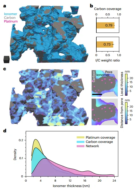
FigureLow-temperature ET reconstruction and ionomer network analysis of microdissected 3M ionomer-HSC-platinum catalyst layers
(3) Platinum-related morphology analysis in the catalyst layer
The authors used the reconstructed volume to further study the morphology and interaction of platinum nanoparticles with carbon supports and ionomer networks. quite a part(46%) of the nanoparticles were found to exist in the nanopores of the carbon primary particles. The average diameters of the inner and outer particles were 2.7 and 3.0 nm, respectively, while the distribution of the outer particles was slightly wider. This may be due to the internal porosity of the carbon support during the synthesis process. Limit the growth of particles. Focusing on the external platinum particles and their connectivity to the ion network, the surface area calculated from the chromatogram was then compared with the volume measurement. The results show that all platinum can be stripped by CO at high relative humidity levels.
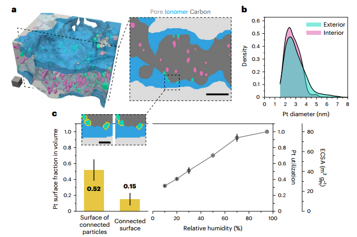
FigureMicro-cuttingAnalysis of platinum-related morphology in 3M ionomer-HSC-platinum catalyst layer
References:
Girod, R., Lazaridis, T., Gasteiger, H.A. et al. Three-dimensional nanoimaging of fuel cell catalyst layers. Nat Catal (2023). https://doi.org/10.1038/s41929-023-00947-y
Artikel sebelumnya
Informasi terkait


Hubungi kami
Situs web: Website
Alamat: No. 66 jalan selatan Xinyuan, distrik Haicang, Kota Xiamen, Provinsi Fujian, Tiongkok

Kode QR akun resmi